Dermatofibroma
A firm benign nodule diagnosed with certainty and removed cleanly.
A dermatofibroma is a firm, sometimes pigmented nodule that sits within the dermis, most often on the lower legs. It is benign, and it characteristically dimples inward when pinched.
- Local
- Anaesthesia
- 30 min
- Duration
- 10–14 days
- Sutures out
- Always
- Histology
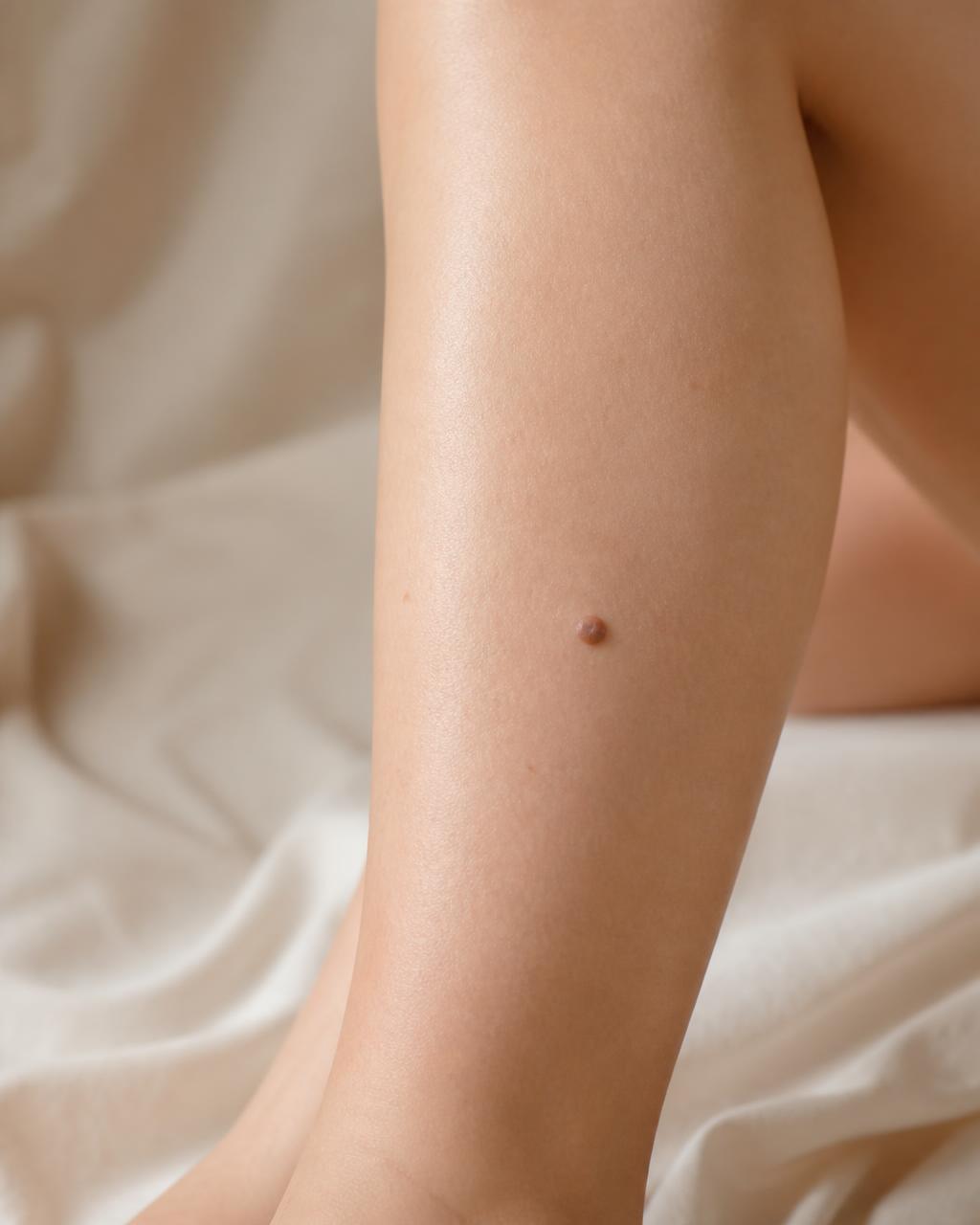
- Dermatoscopic evaluation
- Elliptical excision
- Layered closure
- Histopathology
Diagnosed first, treated second.
Diagnosis matters more than treatment here, because pigmented dermatofibromas can resemble melanoma and rare fibrous tumours can resemble dermatofibromas. Where there is doubt, excision with histology settles it definitively.
- 01Dermatoscopic evaluation
- 02Elliptical excision
- 03Layered closure
- 04Histopathology
- 05Scar management
- A firm nodule on the lower leg
- A pigmented lesion needing diagnosis
- A nodule that is tender or catches when shaving
- A lesion that has changed in appearance
- Cosmetic concern about a visible bump
How the treatment works
A sequence, not a single appointment — each step informs the next.
- 01
Dermatoscopy
The characteristic central white patch and pigment network are assessed.
- 02
Decision
Observation for classic lesions; excision where symptomatic, changing or uncertain.
- 03
Excision
Full-thickness removal under local anaesthesia with careful closure.
- 04
Histology
Pathology confirms the diagnosis and excludes fibrous malignancy.

Results measured, not promised.
What to expect
- 01Diagnosis confirmed with certainty
- 02Lesion removed completely
- 03Tenderness or catching resolved
- 04Melanoma reliably excluded
- 05Well-planned scar on a difficult site
Begin with a diagnosis,
not a guess.
Share a few details and the clinic will reply with available consultation times for dermatofibroma — one-to-one with Dr. Arely.
- Response
- Within 24 hours
- Consultation
- One-to-one, 45 min
- Assessment
- Trichoscopy included
- Location
- Dr. Arely Clinic
Frequently asked
No, it is benign — but pigmented ones are examined carefully to exclude melanoma.
Not necessarily. Removal is for symptoms, cosmetic concern or diagnostic certainty.
Circulation and skin tension on the leg slow healing; closure and aftercare account for that.
Rarely after full-thickness excision.